Vanessa Schoeppler1, Robert Lemanis, Elke Reich1, Tamás Pusztai2, László Gránásy2,3, Igor Zlotnikov1
1B CUBE - Center for Molecular Bioengineering, Technische Universität Dresden, Germany
2Institute for Solid State Physics and Optics, Wigner Research Centre for Physics, P.O. Box 49, Budapest H-1525, Hungary
3BCAST, Brunel University, Uxbridge, Middlesex, UB8 3PH, United Kingdom
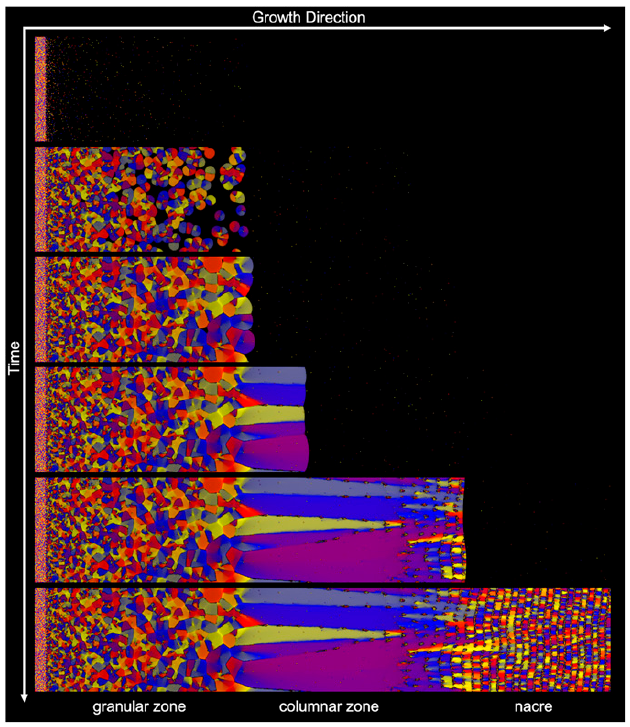
Molluscan shells are a classic model system to study formation–structure–function relationships in biological materials and the process of biomineralized tissue morphogenesis. Typically, each shell consists of a number of highly mineralized ultrastructures, each characterized by a specific 3D mineral–organic architecture. Surprisingly, in some cases, despite the lack of a mutual biochemical toolkit for biomineralization or evidence of homology, shells from different independently evolved species contain similar ultrastructural motifs. In the present study, using a recently developed physical framework, which is based on an analogy to the process of directional solidification and simulated by phase-field modeling, we compare the process of ultrastructural morphogenesis of shells from 3 major molluscan classes: A bivalve Unio pictorum, a cephalopod Nautilus pompilius, and a gastropod Haliotis asinina. We demonstrate that the fabrication of these tissues is guided by the organisms by regulating the chemical and physical boundary conditions that control the growth kinetics of the mineral phase. This biomineralization concept is postulated to act as an architectural constraint on the evolution of molluscan shells by defining a morphospace of possible shell ultrastructures that is bounded by the thermodynamics and kinetics of crystal growth.


